| Home | E-Submission | Sitemap | Contact Us |  
top_img
Environ Health Toxicol > Volume 33:2018 > Article
Hwang, Choi, Kim, Gim, Ha, and Kim: Health effects from exposure to dental diagnostic X-ray

Abstract

The purpose of this review is to summarize the results of studies on of the association between exposure to dental X-rays and health risk. To perform the systematic review, We searched the PUBMED, EMBASE, and MEDLINE databases for papers published before December 15, 2016. A total of 2 158 studies, excluding duplicate studies, were found. Two reviewers independently evaluated the eligibility of each study. The final 21 studies were selected after application of exclusion criteria. In terms of health outcomes, there were 10 studies about brain tumors, 5 about thyroid cancer, 3 about tumors of head and neck areas, and 3 related to systemic health. In brain tumor studies, the association between dental X-ray exposure and meningioma was statistically significant in 5 of the 7 studies. In 4 of the 5 thyroid-related studies, there was a significant correlation with dental diagnostic X-rays. In studies on head and neck areas, tumors included laryngeal, parotid gland, and salivary gland cancers. There was also a statistically significant correlation between full-mouth X-rays and salivary gland cancer, but not parotid gland cancer. Health outcomes such as leukemia, low birth weight, cataracts, and thumb carcinomas were also reported. In a few studies examining health effects related to dental X-ray exposure, possibly increased risks of meningioma and thyroid cancer were suggested. More studies with a large population and prospective design are needed to elaborate these associations further.

INTRODUCTION

Dental diagnostic X-rays are an essential part of dental practice. Although radiation doses have been reduced due to the development of digital techniques, dental diagnostic X-ray imaging remains one of the most common types of radiological procedures that are frequently performed in dental clinics for oral examinations [1,2]. Dental diagnostic X-rays for certain types of examinations, including bitewings, full-mouth series, and panoramic views, are in common use. Considering the lifetime frequency of exposure to dental diagnostic X-rays, even a slight increase in health risk would be of considerable public health importance [3].
Dental X-rays expose patients to relatively low radiation doses. However, the survivors of the Hiroshima atomic bombings provide evidence of increased cancer risk from low doses of radiation [4]. Repeated exposure could also increase cancer risk [5]. Exposure to dental X-rays is associated with potential risk of cancer, which was revealed in previous studies [6,7].
In the head and neck region, cancer risks caused by exposure to dental X-rays have been discussed. Although many epidemiological studies have reported on the association between exposure to dental X-rays and meningioma risk, it is still controversial, as some studies have shown a lack of association [8-10]. A meta-analysis study proposed that there is no clear evidence of a significant association between exposure to dental diagnostic X-rays and the risk of developing meningioma [11]. Several studies have found an association between dental X-ray exposure and increased risks of brain cancer [12,13], tumors of the parotid gland [14] and breast cancer [15] and thyroid cancer [16,17]. In particular, thyroid cancer is one of the most common cancers in the worldwide, and the side effects from dental radiation exposure are likely to contribute to its incidence due to the location of the thyroid gland. Repeated exposure to dental X-rays may result in various health problems including head and neck tumors and various systemic problems. Thus, we conducted a systematic review of papers that reported an association between dental X-ray exposure and overall health risks because no previous reports have summarized these associations.

MATERIALS AND METHODS

The patient, intervention, comparator, outcomes (PICO) method was followed as a viable tool for the systematic review process [18]. The PICO method for this systematic review was as follows. The “P” referred to all patients, the “I” referred to dental X-ray exposure, the “C” referred to dental X-ray non-exposure, and the “O” referred to brain cancer, meningioma, thyroid cancer, leukemia, and other cancers.

Search strategy and data sources

We searched the PUBMED, EMBASE, and MEDLINE databases and performed a manual search for papers. The databases were searched for all related paper published before December 2016. The paper published in all languages were selected. The following search terms were employed.
(i) PUBMED: (“radiography, dental” [MeSH Terms] OR (“radiography” [All Fields] AND “dental” [All Fields]) OR “dental radiography” [All Fields] OR “dental x ray” [All Fields]) AND exposure [All Fields] AND (“brain neoplasms” [MeSH Terms] OR (“brain” [All Fields] AND “neoplasms” [All Fields]) OR “brain neoplasms” [All Fields] OR (“brain” [All Fields] AND “cancer” [All Fields]) OR “brain cancer” [All Fields]); (ii) EMBASE: ‘dental’/exp OR dental AND (‘x ray’/exp OR ‘x ray’) AND (‘exposure’/exp OR exposure); (iii) Manual search was carried out using the reference lists of papers included in the systematic review, on review papers about overall health.

Study inclusion and exclusion

Two reviewers (S.Y.H, E.S.C.) independently assessed the eligibility of each study through the databases based on the predetermined selection criteria. Any disagreements were resolved through discussion. The inclusion criteria were as follows: (i) human study; (ii) health problems including cancers related to dental radiation exposure; (iii) the full text of the study was available. The exclusion criteria were as follows: (i) Radiation dose assessment study; (ii) Radiation safety management study; (iii) Review articles; (iv) Letter and recommendation.

Data extraction

Two authors (M.A.H. and H.Y.K) independently collected the following information: first author’s name, year of publication, study design, dental diagnostic X-ray type, health outcome, significance, risk estimates, and their confidence intervals (CIs). We tried to summarize the significance of test results according to the types of health outcomes and types of the dental x-ray. However, test results in some studies were expressed for more detailed categories such as age groups or frequencies of exposure and occurred either consistent which means that all test were significant or nonsignificant, or inconsistent which means that both coexisted. To solve the problem, the integrated significance was marked as having partial significance (PS) when only some of the characteristics were statistically significant, as having significance (S) when all the characteristics were significant, and as not having significance (NS) when all were nonsignificant.

Quality assessment

We assessed the methodological quality of each study using the Newcastle-Ottawa scale (NOS) [19] which uses a star rating system. A full score is 9 stars, and a score range 5 to 9 stars is considered to be a high methodological quality while a score range 0 to 4 is considered to be poor quality. Nineteen case-control studies and one cohort study were assessed on the quality excluding one case-report study.

RESULTS

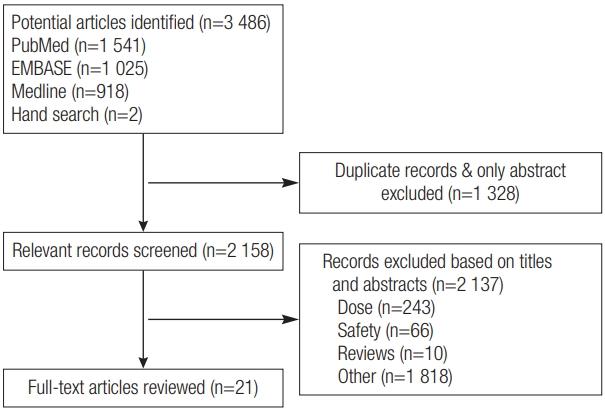
A total of 2 158 studies, except for duplicate studies, were initially collected. The abstracts and titles of the 2 158 papers were assessed. The full-text of the final 21 studies that were selected through classifications of exclusion criteria were read (Figure 1). We assessed information provided by the reporting of case–controls (n=19), cohort studies (n=1), and case studies (n=1). These studies were published between 1997 and 2015. These studies were conducted in the following countries: the United States (n=12), Sweden (n=4), Taiwan (n=1), Australia (n=1), Kuwait (n=1), Japan (n=1), and Syria (n=1).

Quality assessment

The quality of all included studies was summarized in Table 1. The median NOS score of the eligible studies was 4.0, 5.0, and 3.5 for meningioma and tumors in head and neck areas, thyroid cancer, and systemic health outcomes, respectively.

Health-related outcomes

Brain tumors

Table 2 shows 10 research papers on dental diagnostic X-rays and brain tumors. All were case–control studies. Five categorized or specified the types of dental diagnostic X-rays, while the other five did not. Two of these studies also included the panorama and bitewing types of examinations. Detailed analyses according to age groups or exposure frequencies were performed in five studies examining the correlation between the full-mouth examination type and brain tumors.
There were seven papers on meningiomas, three on gliomas, one on acoustic neuroma, and one on vestibular schwannoma, while two papers mentioned unspecific brain cancer. Seven papers reported a significant positive association and three presented a partially positive significant association, while eight had nonsignificant results. One paper reported a negative association between full-mouth X-ray exposure and glioma.
The association with meningioma was significant in 5 of 7 studies, while it was nonsignificant in the remaining 2 studies. Inconsistent results were reported on the association with glioma. A strong positive association was found in relation with vestibular schwannoma, while not with acoustic neuroma. The full-mouth examination type increased the risk of brain tumors significantly in three of the four studies.

Thyroid cancer

There were five research findings related to thyroid cancer (Table 3). As far as research design was concerned, four were case–control studies, while one was a cohort study. The five studies did not specify dental diagnostic X-ray types. In four of the five studies, there were significant correlations between dental diagnostic X-rays and thyroid cancer. One of these studies categorized the subjects by occupation and confirmed the correlation between dental practice and thyroid cancer.

Tumors in head and necks areas

Table 3 lists research findings on the tumors in head and necks areas. Tumors included laryngeal, parotid gland, and salivary gland cancers. As exposure to dental diagnostic X-rays increased, the risk of laryngeal cancer also increased. There was also a statistically significant correlation between full-mouth X-rays and salivary gland cancer, but not parotid gland cancer.

Systemic health outcomes

Leukemia and low birth weight have been reported as systemic health outcomes related to dental X-ray exposure (Table 3). One study categorized dental diagnostic X-ray types and examined correlations with low birth weight (LBW) and showed that only panoramic examination types had a statistically significant correlation with LBW. The risk of leukemia significantly increased in accordance with dental diagnostic X-ray exposure. A case report showed that thumb carcinoma occurred in radiographic technician who had performed dental diagnostic X-ray examinations for 15 years.

DISCUSSION

Patients are certainly exposed to dental diagnostic X-rays, and dental practitioners also potentially receive exposure. Although the level of exposure is lower than that of medical radiation, there is an innate risk from radiation exposure that cannot be ignored. However, there are a few studies on low-dose dental diagnostic X-ray exposure and health effects, except with respect to some parts of the body. In addition, papers report inconsistent statistical significances on dental diagnostic X-ray exposure and health effects, depending on the subjects and research design; therefore, related studies should be systematically organized. Hence, through a systematic literature review, this study included various studies with different research designs and examined the health risks associated with dental diagnostic X-ray exposure. For the literature review, this study examined twenty-one papers on dental diagnostic X-ray exposure and health effects.
Among twenty-one papers that reported a correlation between dental diagnostic X-rays and overall health, eighteen papers assessed a correlation with head and neck areas. The selected papers included ten on brain cancer, five on thyroid cancer, and three on head and neck areas other than the brain and thyroid. Since the oral cavity is anatomically located near the head and neck [36], dental X-ray examinations seem to affect the brain and neck areas. The correlation of X-rays with brain and thyroid cancers has been reported for many decades and seems to explain this finding.
Papers that reported a correlation between dental diagnostic X-ray exposure and overall health of dental practitioners were identified. The present study identified two papers on occupational groups [17,35]. According to a study on dental practitioners and thyroid cancer the risk of thyroid cancer was 13.1 times (95% CI 2.1–389) higher among female dentists and dental hygienists [17]. It is believed that women are more likely to have to thyroid cancer than men due to their hormones [37]. In a case report, a dental radiologist was exposed to dental diagnostic X-rays for 15 years and ended up developing finger cancer, which demonstrates that long-term exposure to low doses of dental diagnostic X-rays can pose a risk to health [35]. Another paper also reported that the cumulative dose for dentists who had worked for a long time was high [38]. Hence, dental practitioners should be aware of exposure to low doses of dental diagnostic X-rays, and risk of accumulative exposure to low-dose radiation from dental X-rays cannot be ruled out.
Dental diagnostic X-ray types were specifically categorized. Especially, panorama examination types are more commonly used than simple dental X-ray types, because panoramic diagnostic information coverage exceeds that of dental diagnostic X-rays [39]. Panoramic examinations also require fewer steps than conventional full-mouth examination types [40]. The full-mouth type had a statistical correlation with meningioma and salivary gland cancers. Meningioma cancer showed four times higher risk at younger ages (<20 years). Unlike adults, children are much more sensitive to radiation exposure due to active cell division [41]. In addition, the panorama examination type showed a correlation with meningioma cancer at higher exposure frequencies. Accordingly, a study on the amount of radiation exposure from each dental diagnostic X-ray type is needed. Nonetheless, the selected papers on thyroid cancer, which has a high level of radiation sensitivity [42], did not categorize dental diagnostic X-ray types, so this could not be examined.
There were some limitations of this study. This study did not specifically categorize exposure measurements, including exposure dose (mGy) and frequency, because each study has diverse radiation exposure categories. This study performed the only systematic review, not was extended to a meta-analysis, because the types of health outcomes were too diverse. However, we could identify trends in the dental diagnostic X-ray studies via this systematic literature review. This study has some important contributions. First, unlike existing literature reviews, this study covered diverse health outcomes as well as meningioma. Second, this study showed evidence that the increased risk of head and neck cancer due to exposure to low doses of dental diagnostic X-rays cannot be ignored.
It should also be noted that no studies have examined the specific types of dental X-rays and prevalence of thyroid cancer, which should be explored in further studies. Further studies are also needed to investigate the health effects of dental diagnostic X-rays in dental practitioners, who may be frequently exposed to high levels of radiation exposure.

REFERENCES

1. Han GS, Cheng JG, Li G, Ma XC. Shielding effect of thyroid collar for digital panoramic radiography. Dentomaxillofac Radiol 2013;42(9):20130265.
crossref pmid pmc
2. Toossi MTB, Akbari F, Roodi SB. Radiation exposure to critical organs in panoramic dental examination. Acta Med Iran 2012;50(12):809-813.
pmid
3. Crane GD, Abbott PV. Radiation shielding in dentistry: an update. Aust Dent J 2016;61(3):277-281.
crossref pmid
4. Preston D, Ron E, Tokuoka S, Funamoto S, Nishi N, Soda M, et al. Solid cancer incidence in atomic bomb survivors: 1958–1998. Radiat Res 2007;168(1):1-64.
crossref pmid
5. Zielinski JM, Ashmore PJ, Band PR, Jiang H, Shilnikova NS, Tait VK, et al. Low dose ionizing radiation exposure and cardiovascular disease mortality: cohort study based on Canadian national dose registry of radiation workers. Int J Occup Med Environ Health 2009;22(1):27-33.
crossref pmid pdf
6. Hall EJ, Brenner DJ. Cancer risks from diagnostic radiology. Br J Radiol 2008;81(965):362-378.
crossref pmid
7. Brenner DJ, Doll R, Goodhead DT, Hall EJ, Land CE, Little JB, et al. Cancer risks attributable to low doses of ionizing radiation: assessing what we really know. Proc Natl Acad Sci USA 2003;100(24):13761-13766.
crossref pmid
8. Burch JD, Craib KJ, Choi BC, Miller AB, Risch HA, Howe GR. An exploratory case-control study of brain tumors in adults. J Natl Cancer Inst 1987;78(4):601-609.
pmid
9. Preston-Martin S, White SC. Brain and salivary gland tumors related to prior dental radiography: implications for current practice. J Am Dent Assoc 1990;120(2):151-158.
crossref pmid
10. Ryan P, Lee MW, North B, McMichael AJ. Amalgam fillings, diagnostic dental x-rays and tumours of the brain and meninges. Eur J Cancer B Oral Oncol 1992;28B(2):91-95.
crossref pmid
11. Xu P, Luo H, Huang G-L, Yin X-H, Luo S-Y, Song J-K. Exposure to Ionizing Radiation during Dental X-Rays Is Not Associated with Risk of Developing Meningioma: A Meta-Analysis Based on Seven Case-Control Studies. PLOS one 2015;10(2):e0113210.
crossref pmid pmc
12. Preston-Martin S, Mack W, Henderson BE. Risk factors for gliomas and meningiomas in males in Los Angeles County. Cancer Res 1989;49(21):6137-6143.
pmid
13. Neuberger JS, Brownson RC, Morantz RA, Chin TD. Association of brain cancer with dental X-rays and occupation in Missouri. Cancer Detect Prev 1991;15(1):31-34.
pmid
14. Preston-Martin S, Henderson BE, Bernstein L. Medical and dental X rays as risk factors for recently diagnosed tumors of the head. Natl Cancer Inst Monogr 1985;69: 175-179.
pmid
15. Ma H, Hill CK, Bernstein L, Ursin G. Low-dose medical radiation exposure and breast cancer risk in women under age 50 years overall and by estrogen and progesterone receptor status: results from a case–control and a case–case comparison. Breast Cancer Res Treat 2008;109(1):77-90.
crossref pmid
16. Memon A, Godward S, Williams D, Siddique I, Al-Saleh K. Dental x-rays and the risk of thyroid cancer: a case-control study. Acta Oncol 2010;49(4):447-453.
crossref pmid
17. Wingren G, Hallquist A, Hardell L. Diagnostic X-ray exposure and female papillary thyroid cancer: a pooled analysis of two Swedish studies. Eur J Cancer Prev 1997;6(6):550-556.
crossref pmid
18. Bae JM. An overview of systematic reviews of diagnostic tests accuracy. Epidemiol Health 2014;36: e2014016.
crossref pmid pmc pdf
19. Stang A. Critical evaluation of the Newcastle-Ottawa scale for the assessment of the quality of nonrandomized studies in meta-analyses. Eur J Epidemiol 2010;25(9):603-605.
crossref pmid
20. Preston-Martin S, Paganini-Hill A, Henderson BE, Pike MC, Wood C. Case-control study of intracranial meningiomas in women in Los Angeles County, California. J Natl Cancer Inst 1980;65(1):67-73.
pmid
21. Preston-Martin S, Mimi CY, Henderson BE, Roberts C. Risk factors for meningiomas in men in Los Angeles County. J Natl Cancer Inst 1983;70(5):863-866.
pmid
22. Rodvall Y, Ahlbom A, Pershagen G, Nylander M, Spannare B. Dental radiography after age 25 years, amalgam fillings and tumours of the central nervous system. Oral Oncol 1998;34(4):265-269.
crossref pmid
23. Longstreth W, Phillips LE, Drangsholt M, Koepsell TD, Custer BS, Gehrels JA, et al. Dental X‐rays and the risk of intracranial meningioma. Cancer 2004;100(5):1026-1034.
crossref pmid
24. Claus EB, Calvocoressi L, Bondy ML, Schildkraut JM, Wiemels JL, Wrensch M. Dental x‐rays and risk of meningioma. Cancer 2012;118(18):4530-4537.
crossref pmid pmc
25. Han Y-Y, Berkowitz O, Talbott E, Kondziolka D, Donovan M, Lunsford LD. Are frequent dental x-ray examinations associated with increased risk of vestibular schwannoma? Clinical article. J Neurosurg 2012;117(Special Suppl):78-83.
crossref
26. Lin M-C, Lee C, Lin C, Wu Y, Wang H, Chen C, et al. Dental diagnostic X-ray exposure and risk of benign and malignant brain tumors. Ann Oncol 2013;24(6):1675-1679.
crossref pmid pdf
27. Wingren G, Hatschek T, Axelson O. Determinants of papillary cancer of the thyroid. Am J Epidemiol 1993;138(7):482-491.
crossref pmid pdf
28. Hallquist A, Hardell L, Degerman A, Wingren G, Boquist L. Medical diagnostic and therapeutic ionizing radiation and the risk for thyroid cancer: a case-control study. Eur J C Prev 1994;3(3):259-267.
crossref
29. Neta G, Rajaraman P, Berrington de Gonzalez A, Doody MM, Alexander BH, Preston D, et al. A prospective study of medical diagnostic radiography and risk of thyroid cancer. Am J Epidemiol 2013;177(8):800-809.
crossref pmid pmc pdf
30. Hinds M. Anatomic distribution of malignant melanoma of the skin among non-Caucasians in Hawaii. Br J Cancer 1979;40(3):497-499.
crossref pmid pmc pdf
31. Preston-Martin S, Thomas DC, White SC, Cohen D. Prior exposure to medical and dental x-rays related to tumors of the parotid gland. J Natl Cancer Inst 1988;80(12):943-949.
crossref pmid pdf
32. Horn-Ross PL, Ljung BM, Morrow M. Environmental factors and the risk of salivary gland cancer. Epidemiology 1997;8(4):414-419.
crossref pmid
33. Nishi M, Miyake H. A case-control study of non-T cell acute lymphoblastic leukaemia of children in Hokkaido, Japan. J Epidemiol Community Health 1989;43(4):352-355.
crossref pmid pmc
34. Hujoel PP, Bollen A-M, Noonan CJ, del Aguila MA. Antepartum dental radiography and infant low birth weight. JAMA 2004;291(16):1987-1993.
crossref pmid
35. Halboub ES, Barngkgei I, Alsabbagh O, Hamadah O. Radiation-induced thumbs carcinoma due to practicing dental X-ray. Contemp Clin Dent 2015;6(1):116-118.
crossref pmid pmc
36. Vissink A, Jansma J, Spijkervet F, Burlage F, Coppes R. Oral sequelae of head and neck radiotherapy. Crit Rev Oral Biol Med 2003;14(3):199-212.
crossref pmid
37. Shore RE. Issues and epidemiological evidence regarding radiationinduced thyroid cancer. Radiat Res 1992;131(1):98-111.
crossref pmid
38. Kim YJ, Cha ES, Lee WJ. Occupational radiation procedures and doses in South Korean dentists. Community Dent Oral Epidemiol 2016;44(5):476-484.
crossref pmid
39. Horton PS, Sippy FH, Kerber PE, Paule CL. Analysis of interpretations of full-mouth and panoramic surveys. Oral Surg Oral Med Oral Pathol 1977;44(3):468-475.
crossref pmid
40. Rushton V, Horner K. The use of panoramic radiology in dental practice. J Dent 1996;24(3):185-201.
crossref pmid
41. Myers DR, Shoaf HK, Wege WR, Carlton WH, Gilbert MA. Radiation exposure during panoramic radiography in children. Oral Surg Oral Med Oral Pathol 1978;46(4):588-593.
crossref pmid
42. Hiraoka T, Miller RC, Gould MN, Kopecky KJ, Ezaki H, Takeichi N, et al. Survival of human normal thyroid cells after X-ray irradiation. Int J Radiat Biol Relat Stud Phys Chem Med 1985;47(3):299-307.
crossref pmid

Figure 1.
Flow chart of identification of eligible studies to final inclusion.
eht-33-4-e2018017f1.jpg
Table 1.
Newcastle-Ottawa Scale Quality Assessment of included studies (N=20)
Type Author(year) Study design Selection Comparability Exposure Total
Brain tumor Preston-Martin (1980) Case-control 2 1 1 4
Preston-Martin (1983) Case-control 2 1 1 4
Preston-Martin (1989) Case-control 2 1 1 4
Neuberger JS (1991) Case-control 2 1 1 4
Ryan P (1992) Case-control 2 0 1 3
Rodvall y (1998) Case-control 2 2 1 4
Longstreth w (2004) Case-control 2 2 2 6
Claus E (2012) Case-control 3 2 2 7
Han YY (2012) Case-control 3 2 2 7
Lin MC (2013) Case-control 3 1 1 5
Thyroid cancer Wingren G (1993) Case-control 2 2 1 5
Hallquist A (1994) Case-control 2 1 1 4
Wingren G (1997) Case-control 2 2 1 5
Memon A (2010) Case-control 3 1 2 6
Neta (2013) Prospective cohort* 3 2 2 7
Tumors in head and neck areas Hinds MW (1979) Case-control 2 1 1 4
Preston-Martin S (1988) Case-control 2 1 1 4
Horn-Rose PL (1997) Case-control 2 1 1 4
Systemic health outcome Motoi (1989) Case-control 1 0 0 1
Hujoel P (2004) Case-control 3 1 2 6

* Newcastle-Ottawa Scale Quality Assessment: cohort

Table 2.
Summary of studies on association between dental X-ray exposure experience and brain tumor
Author (year) Country Study design No. of exposed case/control Dental X-ray types Health outcome Significancea
Preston-Martin S (1980) [20] US Case-control 189/185 Full mouth Meningioma PS Strong positive association with early exposure 20 yr old (OR = 4.0, P < 0.01) among women
Nonsignificant with more than five dental X-ray exposures (OR = 1.6, p = 0.14)
Preston-Martin S (1983) [21] US Case-control 120/105 Full mouth Meningioma PS Strong positive association with early exposure 20 yr old (OR = 7.0, P = 0.04) among men
Nonsignificant with five or more dental X-ray exposures before 1945 (OR=2.7, p=0.11)
Preston-Martin S (1989) [12] US Case-control 272/272 Full mouth Meningioma NS -
Full mouth Glioma PS Positive association with exposure after age 25 (0R=1.0, P<0.04)
Neuberger JS (1991) [13] US Case-control 7/25 Any Brain cancer S Strong positive association (OR = 10.66, 95%CI = 1.95-58.28)
Ryan P (1992) [10] Australia Case-control 6/110 Any Meningioma NS -
Any Gliomas S Negative association (OR=0.42, 95%CI=1.95-58.28)
Rodvall Y (1998) [22] Sweden Case-control 66/65 Any Total CNS tumor NS -
24/65 Any Meningioma S Positive association (RR=2.1, 95%CI=1.0-4.3)
34/65 Any Glioma NS -
8/65 Any Acoustic neuroma NS -
Longstreth W (2004) [23] US Case-control 200/400 Full mouth Meningioma S Positive association (OR=2.06, 95%CI=1.03-4.17)
Others (panorama, cephalometric) NS -
Claus E (2012) [24] US Case-control 1 433/1 350 Full mouth Meningioma NS -
Bitewing S Positive association (OR=2.0, 95%CI=1.4-2.9)
Panorama S Positive association (OR=3.0, 95%CI=1.6-5.6) at frequent exposure, yearly or more
Han YY (2012) [25] US Case-control 343/343 Any Vestibular schwannoma S Strong positive association (OR=4.26, 95%CI=1.49-12.18)w
Lin MC (2013) [26] Taiwan Case-control 4 123/16 492 Any Benign Brain tumor S Positive association (OR=1.39)
Any Malignant Brain tumor NS -

a PS: partial significance S: significance NS: no significance

Table 3.
Summary of studies on association between dental X-ray exposure experience and thyroid cancer, tumors in head and neck areas, and other health outcomes
Author (year) Country Study design No. of exposed case/control Dental X-ray types Health outcome Significancea
Thyroid cancer
Wingren G (1993) [27] Sweden Case-control 11/12 Any Papillary thyroid cancer S Positive association (OR = 2.8 ,95%CI = 1.1-7.5)
Hallquist A (1994) [28] Sweden Case-control 14/34 Any Papillary thyroid cancer NS -
Wingren G (1997) [17] Sweden Case-control 7/1 Any Papillary thyroid cancer S Occupation: dentists or dental assistants (OR = 13.1 95%CI: 2.1-389)
Memon A (2010) [16] Kuwait Case-control 313/313 Any Thyroid cancer S Positive association (OR = 2.1, 95%CI: 1.4-3.1)
Neta (2013) [29] US Prospective cohort 251/75 000c Any Thyroid cancer S Positive association (RR = 1.13 95%CI: 1.01-1.26)
Tumors in head and neck areas
Hinds MW (1979) [30] US Case-control 47/47 Any Laryngeal cancer PS Heavy smoker: Positive association (RR = 7.5, P = 0.02) at frequent exposure ≥ 10
Preston-Martin S (1988) [31] US Case-control 408/408 Any Malignant parotid gland tumor NS -
Any Benign parotid gland tumor NS -
Horn-Rose PL (1997) [32] US Case-control 106/122 Full mouth Salivary gland cancer S Positive association (OR = 1.6, 95%CI:1.0-2.7)
Systemic health outcome
Motoi N (1989) [33] Japan Case-control 63/126 Any Leukemia S Positive association (RR = 1.4)
Hujoel P (2004) [34] US Case-control 1 117/4 468 Full mouth Low-birth-weight NS -
Panoramic S Positive association with LBWb and NBWb(p = 0.009)
Bitewings NS -
Esam S (2015) [35] Syria Case report - - Thumbs carcinoma - Dental Radiographer: A dental radiographer developed thumbs carcinoma after 15 years of practicing.

a PS: partial significance S: significance NS: no significance.

b LBW: low birth weight, birth weight <2 500 g; NBW: normal birth weight, birth weight≥2 500 g.

c No. of exposed case/population.

TOOLS
PDF Links  PDF Links
PubReader  PubReader
ePub Link  ePub Link
XML Download  XML Download
Full text via DOI  Full text via DOI
Download Citation  Download Citation
  Print
Share:      
METRICS
23
Crossref
0
Scopus
18,146
View
278
Download
Editorial Office
Division of Environmental Science and Ecological Engineering, Korea University
145 Anam-ro, Seongbuk-gu, Seoul 02841, Korea
Tel : +82-32-560-7520   E-mail: envitoxic@gmail.com
About |  Browse Articles |  Current Issue |  For Authors and Reviewers
Copyright © 2024 by The Korean Society of Environmental Health and Toxicology & Korea Society for Environmental Analysis.     Developed in M2PI